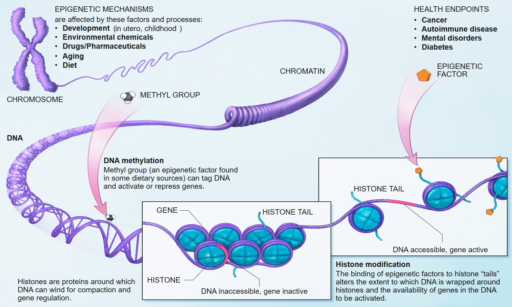
Le sommeil est important pour maintenir la santé du cerveau. Pendant le sommeil, le cerveau élimine les déchets métaboliques, les protéines et les débris cellulaires qui s'accumulent pendant l'activité cérébrale diurne. Ceet élimination se fait via grâce à des systèmes interconnectés d'activité neuronale et de flux de liquide interstitiel et céphalo-rachidien. enter image description here Des études précliniques ont montré que pendant le sommeil, l'infiltration du liquide interstitiel et céphalo-rachidien le long des espaces périvasculaires augmente, augmentant ainsi la clairance des solutés interstitiels. Lorsqu'ils sont agrandis, les espaces périvasculaires sont visibles par imagerie par résonance magnétique cérébrale (IRM). Le volume et le nombre de espaces périvasculaires augmentent avec l'âge. Ce phénomène est associés à des facteurs de risque vasculaire tels que l'hypertension, des marqueurs de microangiopathie telle que la leucoaraiose et entraine des effets néfastes sur la santé. Une question importante est de savoir si le manque de sommeil est la cause de ces risques vasculaires, ou simplement leur conséquence.

La microangiopathie est une angiopathie qui atteint des vaisseaux sanguins de petits calibres. Elle est souvent une complication du diabète et de l'hypertension artérielle. La présence de leucoaraiose sur l'IRM a également été associée à un déclin fonctionnel, à des troubles de la marche et à la dépression, peut-être parce qu'ils perturbent les réseaux neuronaux. enter image description here

Souvent, les leucoaraiose sont dus à un processus appelé infarctus incomplet, c'est à dire une réduction chronique du flux sanguin dans les zones profondes du cerveau causée par l'artériolosclérose, la lipohyalinose ou la nécrose fibrinoïde des petites artères et artérioles cérébrales. Un tel flux sanguin réduit conduit à l'hypoxie, altère les mécanismes d'autorégulation cérébrale et favorise la transcription des gènes inflammatoires, la rupture de la barrière hémato-encéphalique et l'entrée de protéines pro-inflammatoires dans les parois des vaisseaux et le parenchyme cérébral. Même en l'absence d'infarctus franc, ces processus entraînent une démyélinisation, une perte axonale, une réduction de la densité gliale, une vacuolisation et une atrophie du cortex sus-jacent.

En outre, d'autres processus pouvant être impliqués dans la genèse des leucoaraiose comprennent le dysfonctionnement des cellules précurseurs des oligodendrocytes, la défaillance du système glymphatique, la collagénose veineuse. Les études IRM qui utilisent des techniques telles que l'imagerie du tenseur de diffusion et le transfert d'aimantation pour examiner la diffusivité de l'eau et l'intégrité de la substance blanche montrent que certains des changements physiopathologiques précoces se produisent également dans les zones de la substance blanche qui semblent normales sur l'IRM conventionnelle. Ces études IRM des études suggèrent que les leucoaraiose visibles ne sont «que la pointe de l'iceberg» et que la physiopathologie sous-jacente est un processus diffus affectant les petits vaisseaux sanguins dans une grande partie de la substance blanche et d'autres parties du cerveau.

Les marqueurs de la qualité du sommeil ont été associés à plusieurs indicateurs structurels de la détérioration de la santé cérébrale, à savoir les hyperintensités de la substance blanche (Leucoaraiose) d'origine vasculaire présumée, les microhémorragies et la perte de tissu cérébral d'apparence normale, tous évalués à l'aide IRM. Cependant, les études démontrant des associations causales définitives entre le sommeil et la santé des tissus cérébraux au fil du temps chez les personnes âgées vivant dans la communauté font défaut.

Des scientifiques Ecossais essayent de répondre à cette question dans une nouvelle publication, pour cela ils utilisent les données longitudinales des personnes âgées vivant dans la communauté de la Lothian Birth Cohort 1936. Les participants à l'étude sont donc des personnes âgées toutes nées en 1936 à Édimbourg et dans les environs (Lothian). Le Lothian est une région d'Écosse où la langue des Lothiens et de l'ancien royaume de Northumbria est encore pratiquée. La Lothian Birth Cohort 1936 est une vaste étude basée sur la population du vieillissement cognitif, cérébral et général. Les auteurs se sont intéressés à toute association entre la durée, la qualité et l'efficacité du sommeil autodéclarées, la charge espaces périvasculaires au début de la 8e décennie de la vie et la détérioration des volumes cérébraux normaux et anormaux au cours de la décennie, à l'aide de l'IRM structurelle.

Là où des associations existent, les scientifiques ont cherché à savoir si la charge espaces périvasculaires influence ou non l'association entre le sommeil et les changements structurels du cerveau sur une période de temps. Ils ont testé trois hypothèses: (1) les mesures du sommeil serait associées indépendamment à une détérioration ultérieure du tissu cérébral, (2) la charge espaces périvasculaires serait associée à une détérioration du tissu cérébral, (3) l'association entre le sommeil et les changements structurels du cerveau serait en partie médiée par le espaces périvasculaires fardeau.

Dans ce grand échantillon d'individus, le fardeau des espaces périvasculaires élargies visibles dans l'examen IRM du cerveau à l'âge d'environ 73 ans était associé à une aggravation ultérieure de la santé de la substance blanche. Celle-ci était reflétée par une diminution plus rapide du volume de substance blanche d'apparence normale, une plus grande augmentation de leucoaraiose et de la «mesure des dommages à la substance blanche» des années 73 à 79.

Les personnes qui ont signalé une moins bonne efficacité du sommeil et plus de sommeil diurne étaient celles qui ont également connu une baisse significativement plus importante de la santé de leur substance blanche entre 73 et 79 ans, et leurs résultats montrent que cela pourrait être en partie médié par la charge espaces périvasculaires.

Ni la charge espaces périvasculaires, ni la qualité du sommeil ou les indicateurs d'efficacité n'ont été associés à une détérioration du volume de tissue cérébral ou des changements de matière grise au cours de la même période.

Une analyse longitudinale récente à 5 points dans le temps sur 28 ans dans la même cohorte avait auparavant identifié quatre groupes de trajectoires différents avec des durées moyennes de sommeil différentes allant d'environ 5 à 8 h, mais n'avait trouvé aucune différence significative dans la microstructure de la matière blanche ou les volumes de matière grise entre les groupes.

Une autre étude à la fin de l'âge adulte [60–82 ans] avait déjà révélé que le nombre de fois où les participants ont signalé une mauvaise qualité de sommeil sur cinq points temporels couvrant une période de 16 ans avant l'analyse, n'était pas associé à ces mesures de la substance blanche, suggérant ainsi que seule la qualité actuelle du sommeil affectait la substance blanche.

Avec cette étude, les résultats peuvent indiquer soit un effet à court terme du manque d'efficacité du sommeil sur la santé de la substance blanche à la fin de l'âge adulte, soit que l'altération des habitudes de sommeil chez les personnes âgées peut être liée à des changements cérébraux sous-jacents mais ne provoque pas nécessairement les changements cérébraux.

Bien que l'association entre les espaces périvasculaires élargi et le leucoaraiose ait été rapportée, l'étude actuelle suggère qu'en outre, une augmentation des espaces périvasculaires visible (en volume, en nombre ou en scores visuels) prédit également le taux d'atrophie de la substance blanche et la structure de la substance blanche. dommages à la fin de l'âge adulte, mais pas de changements de volume total du cerveau et de la matière grise.

Les troubles du sommeil sont associés à une détérioration plus rapide de la santé de la substance blanche chez les septuagénaires. Une charge plus élevée des espaces périvasculaires est associée à une détérioration plus rapide de la santé de la substance blanche chez les septuagénaires. Les espaces périvasculaires interviennent en partie dans l'effet du sommeil sur la santé du cerveau.

Le principal apport de cette étude semble donc être que c'est la détérioration de la santé vasculaire, plutôt que le manque de sommeil, qui amène la détérioration de la matière blanche.

Cependant, il faut faire preuve de prudence en ce qui concerne le manque apparent d'association avec la matière grise et les taux d'atrophie totale du tissu cérébral. Le logiciel utilisé pour générer des volumes de matière grise, FSL-FAST, bien que considéré comme un étalon-or pour la segmentation des tissus d'apparence normale, ne discrimine pas la matière grise saine de la matière grise malsaine.

L'absence d'association entre les paramètres du sommeil et l'atrophie de la matière grise est moins surprenante.

Une étude précédante examinant la qualité et la quantité du sommeil en relation avec les changements corticaux avait indiqué que la qualité du sommeil et les troubles du sommeil autodéclarés étaient liés à l'amincissement du cortex temporal latéral droit.

Aucune information sur l'apnée du sommeil n'a été recueillie, mais dans les dossiers médicaux, elle n'a été signalée que chez un seul participant.

Quadruple Misfolded Proteins in Older Adults

- Posted by admin in English

In this cohort study, Shama Karanth and colleagues from University of Kentucky, Lexington, investigated quadruple misfolded proteins and other proteinopathy combinations in a cohort of 375 deceased individuals with autopsy data in the University of Kentucky Alzheimer Disease Center (UK-ADC).

At least 3 proteinopathies were observed in 50% of brains.

In addition, quadruple misfolded proteins were associated with severe cognitive impairment at least 12 years before death.

Participants with 3 proteinopathies tended to have poorer global cognition earlier than with the presence of only tau and Aβ and were likely to have higher Braak stages.

Previous studies have reported cognitive decline associated with the presence of mixed pathologies, with study to study differences in methods and proteinopathies, the assessment and inclusion of cerebrovascular pathologies, and hippocampal sclerosis. In the present sample, as in other community based cohorts, FTLD in old age was rare (with an estimated incidence of 8.9 of 100 000 in individuals aged 60 to 69 years; no incidence data are available for older age groups) and was not found in brains of individuals who began follow up with normal cognition.

Individuals with FTLD-TDP-43 with data in the UK-ADC Brain Bank were recruited from a dementia clinic. The scientists excluded 6 individuals with FTLD-TDP-43 in the study; none had the quadruple misfolded proteins phenotype. No discernible overlap in any FTLD feature was observed in these individuals other than presence of TDP-43 proteinopathy, which is now detected in many different neurological diseases outside of the amyotrophic lateral sclerosis–FTLD spectrum.

Cognitive impairment was associated with quadruple misfolded proteins at autopsy, with 89.1% of participants developing dementia and some experiencing profound impairment up to 12 years before death. This finding suggests that quadruple misfolded proteins occur before end stage Alzheimer disease neuropathological change (ie, before high Braak stage).

Consistent with this hypothesis, the mild cognitive impairment to dementia transition was, on average, fastest in the quadruple misfolded proteins group. Estimation of the group cognitive trajectories was aided by the relatively long followup (mean duration of 10.4 years).

These data provide the basis for a novel hypothesis that quadruple misfolded proteins have a more aggressive phenotype from the early stages of the disease rather than accruing additional pathologies only after Alzheimer disease neuropathological change has progressed to high levels. About 10% of these participants died with normal cognition, and previous research has shown quadruple misfolded proteins were present in persons with apparently normal cognition. In the present study, all individuals with quadruple misfolded proteins who had normal cognition at the last visit before death had lower Braak NFT stages (I to III), had no APOEε4 allele, and were predominantly male (4 of 5 participants).

These individuals may represent an early stage of quadruple misfolded proteins, but there are complexities: clinical presentation of proteinopathy combinations may be cohort specific and depend on other currently unknown factors. Older cohorts that survive into advanced old age, like those in the UK-ADC study, may be more likely to experience multiple proteinopathies than younger cohorts. As previously described, APOE appeared to be associated with multiple proteinopathies in this study, particularly those proteinopathy combinations including Aβ plaques. Carriers of APOEε allele not only had increased odds of tau and Aβ, an expected result, but also had higher odds of tau, Aβ, and αsynuclein; tau, Aβ, and TDP-43; and quadruple misfolded proteins.

Unlike previous studies, this study did not find evidence that the ε4 allele was associated with tau or TDP-43 in the absence of Aβ, but the sample size was relatively small. The temporality of protein misfolding may play a clinically important and differentiating role in disease progression.

Autopsy data, although cross-sectional by nature, are compatible with the hypothesis that Aβ aggregates precede, and perhaps stimulate or exacerbate, the widespread misfolding of tau, TDP-43, and α-synuclein. These results suggest that TDP-43 pathology may be associated with poor global cognition.

The presence of multiple proteinopathies, particularly the quadruple misfolded proteins phenotype, appeared to have been associated with the cognitive decline in deceased individuals who participated in a longitudinal community based study at the UK-ADC.

These observations have potentially significant implications for clinical practice and public health, given that strategies to prevent or manage AD dementia may be complicated by the unrecognized presence of multiple additional neuropathologies.

Two epigenetic regulators interfere with healthy aging

- Posted by admin in English

It has long been assumed that lifespan and health are strongly correlated, but although there has been an overall increase in human life expectancy in recent decades, it is too often accompanied by deterioration of health.

A new study published on February 26 in Nature shows the influence of two epigenetic regulators on aging. Scientists led by Jie Yuan from the Chinese Academy of Sciences in Shanghai have studied the BAZ-2 and SET-6 proteins in Caenorhabditis elegans worms, which are orthologs of the human proteins BAZ2B and EHMT1.

Through genome-wide RNA-interference-based screening of genes that regulate behavioral deterioration in aging C. elegans, the researchers identified 59 genes as potential health modulators during aging. Essentially the proteins expressed by these genes, read and write epigenetic signals.

Among these modulators, they found that a neuronal epigenetic reader, BAZ-2, and a neuronal histone SET-6, accelerate the deterioration of the behavior of C. elegans by reducing the mitochondrial function, and repressing the expression of the encoded mitochondrial proteins. in the cell nucleus.

The researchers found that the levels of the two proteins increase with age in C. elegans and mice, which in turn attenuates the expression of genes involved in mitochondrial function.

BAZ-2 and SET-6 are complementary epigenetic mechanisms. SET-6 is an "epigenetic writer" and BAZ-2 is an "epigenetic reader" which recognizes modified histones and recruits transcriptional regulators.

Histones are proteins located in the nucleus of eukaryotic cells. They are the main protein components of chromosomes. They are closely associated with DNA and allow their compaction, but they also modify the expression of proteins by various epigenetic mechanisms known as the "histone code".

enter image description here Source Wikipedia.

How do BAZ-2 and SET-6 accelerate aging? The researchers found that the two proteins bind together to the promoter regions of more than 2,000 genes, and decrease their expression via methylation of histones. Among these target genes are many mitochondrial genes encoded nuclear. By suppressing the expression of these genes, BAZ-2 and SET-6 reduce oxygen consumption and ATP production, and decrease the critical stress responses that maintain mitochondrial proteostasis. The resulting metabolic slowdown discourages the worms from assimilating their food and they mate less.

This mechanism is conserved in the neurons of cultured mice and human cells. What about the orthologs of these epigenetic proteins in humans? A review of the databases shows that expression by human orthologs of the two proteins mentioned above, BAZ2B and EHMT1, increases with age and is positively correlated with the progression of Alzheimer's disease. Researchers have verified that ablation of BAZ-2 mouse ortholog Baz2b attenuates age-dependent body weight gain and prevents cognitive decline in aging mice.

enter image description here 
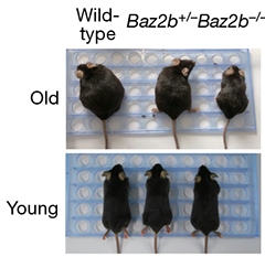
While wild-type mice grew fat with age, animals lacking both copies of the epigenetic reader Baz2b stayed trim, indicating improved mitochondrial function. [Yuan et al., Nature, 2020.]

However, it must be asked whether BAZ-2 and SET-6 would rather mediate age-related physiological adaptation, rather than the agents of aging itself. Indeed their action could reflect a mechanism of adaptation to a progressively more hostile biological environment.

Old Article 5

- Posted by system in English

Old Article 4

- Posted by system in English

Old Article 3

- Posted by system in English

Old Article 2

- Posted by system in English

Old Article 1

- Posted by system in English

Analysis of the Text: Significance, Importance, Timeliness, and Relevance

The text discusses the neural mechanisms underlying the difficulty in turning while walking in individuals with Parkinson's disease (PD), a common symptom that leads to falls and loss of independence. The study examines the activity in the motor cortex and basal ganglia using implanted devices that record and stimulate the brain.

Significance: The topic is significant because it addresses a critical aspect of PD management, which is the prevention of falls and maintenance of independence. Falls are a leading cause of morbidity and mortality in individuals with PD, and understanding the neural mechanisms behind this difficulty can lead to more effective interventions.

Importance: The importance of this study lies in its potential to establish circuit targets for adaptive brain stimulation, a non-invasive therapy that can improve turning and reduce falls in individuals with PD. This can have significant implications for disease management and quality of life.

Timeliness: The study's focus on PD is particularly timely, given the growing awareness of the need for more effective and non-invasive treatments for this condition. PD affects over 10 million people worldwide, and the development of more targeted therapies is crucial to improving patient outcomes.

Relevance: The study is relevant to the broader field of motor control and neurology, as it sheds light on the complex neural interactions underlying human movement. The findings have implications for our understanding of PD and potentially other movement disorders.

Insights into the Relationship between Items in the Text:

  • Motor cortex and basal ganglia interactions: The study highlights the importance of dynamic interactions between these brain regions in enabling complex movement, particularly turning.
  • Beta-band activity and synchrony: The text shows that excessive beta synchrony is associated with impaired turns, whereas reduced beta-band activity is linked to successful turns.
  • Medication and deep brain stimulation: The study demonstrates that these treatments improve turning through distinct circuit mechanisms, involving dopamine suppression of abnormal pallidal beta activity and stimulation restoration of cortical flexibility.

Usefulness for Disease Management or Drug Discovery:

The study provides valuable insights into the neural mechanisms underlying the difficulty in turning while walking in individuals with PD. The findings suggest that adaptive brain stimulation can be an effective therapy for reducing falls and improving independence in individuals with PD. This can lead to the development of more targeted and non-invasive treatments for this condition.

Original Information Beyond the Obvious:

While the study does not provide entirely new information, it offers a nuanced understanding of the neural mechanisms underlying PD, highlighting the importance of dynamic cortical-basal ganglia interactions in enabling complex movement. The findings also suggest that medication and deep brain stimulation can improve turning through distinct circuit mechanisms, which is a valuable addition to our understanding of PD treatment.

Comparison with the State of the Art:

The study contributes to our understanding of PD by providing a more detailed understanding of the neural mechanisms underlying the difficulty in turning while walking. This knowledge can be used to develop more effective and targeted treatments for this condition, building on existing research in the field of PD treatment and motor control.

In conclusion, the study provides significant insights into the neural mechanisms underlying the difficulty in turning while walking in individuals with PD, highlighting the importance of dynamic cortical-basal ganglia interactions and the potential of adaptive brain stimulation as a non-invasive therapy. While not providing entirely new information, the study builds on existing research and offers a valuable contribution to our understanding of PD and its treatment.

Read the original article on medRxiv

Significance of the Topic

The topic of this text revolves around understanding the molecular mechanisms underlying the genetic risks associated with Alzheimer's disease (Alzheimer's disease) conferred by the Apolipoprotein E (APOE) gene. Specifically, it explores the role of the 4 and 2 alleles of the APOE gene in modulating Alzheimer's disease pathology. The significance of this topic lies in its potential to shed light on the underlying biology of Alzheimer's disease, a complex and multifactorial disease.

Importance

Alzheimer's disease is a devastating and debilitating neurodegenerative disorder that affects millions of people worldwide. Understanding the genetic and molecular mechanisms underlying Alzheimer's disease is crucial for developing effective therapeutic strategies and preventing or slowing disease progression. The APOE gene has been associated with Alzheimer's disease risk, with the 4 allele being a well-established risk factor.

However, the underlying molecular mechanisms remain poorly characterized, making this research area highly important and timely.

Timeliness

The text is timely as it addresses a critical knowledge gap in the field of Alzheimer's disease research. Recent advances in proteomics and genomics have enabled researchers to systematically profile APOE-associated proteomic alterations in human samples, providing new insights into the molecular mechanisms underlying Alzheimer's disease. The text leverages these advances to investigate the role of the APOE 4 and 2 alleles in Alzheimer's disease pathology, making it a timely contribution to the field.

Relevance

The text has significant relevance to Alzheimer's disease research, as it provides novel insights into the molecular mechanisms underlying APOE-driven Alzheimer's disease pathology. The findings have implications for the development of therapeutic strategies for early intervention and potentially for the identification of new targets for Alzheimer's disease treatment.

Furthermore, the text highlights the importance of considering the APOE 4 and 2 alleles as distinct risk factors for Alzheimer's disease, rather than just focusing on the 4 allele.

Analysis of the Text

The text presents a comprehensive analysis of APOE-associated proteomic alterations across five cohorts, using a range of proteomics platforms and samples, including plasma and cerebrospinal fluid (cerebrospinal fluid). The study uses systematic profiling to identify a comprehensive APOE-protein network and applies mediation modeling to classify genotype-related signals as upstream mediators, downstream consequences, or APOE-specific changes. The text then leverages cerebrospinal fluid beta-amyloid (A ) biomarker data to improve temporal resolution and isolate early, A -independent proteomic programs.

The findings of the text are significant, as they provide novel insights into the molecular mechanisms underlying APOE-driven Alzheimer's disease pathology. The study identifies allele-specific, temporally structured proteomic signatures that precede Alzheimer's disease pathology, offering potential therapeutic targets for early intervention. The text highlights the importance of considering the APOE 4 and 2 alleles as distinct risk factors for Alzheimer's disease and underscores the challenges in reproducibility associated with proteomics studies.

Usefulness for Disease Management and Drug Discovery

The text provides valuable insights for Alzheimer's disease disease management and drug discovery, as it highlights the potential therapeutic targets for early intervention. The identification of allele-specific, temporally structured proteomic signatures offers a new perspective on Alzheimer's disease pathology and provides a starting point for the development of novel therapeutic strategies. The text also underscores the importance of considering individual variability in APOE genotype as a critical factor in Alzheimer's disease risk, which may inform personalized medicine approaches.

Original Information Beyond the Obvious

While the text presents novel insights into the molecular mechanisms underlying APOE-driven Alzheimer's disease pathology, it does not break new ground in terms of fundamental understanding.

However, the systematic profiling and mediation modeling approaches used in the study provide a comprehensive and nuanced understanding of APOE-associated proteomic alterations, which is a significant advance in the field. The text highlights the challenges associated with reproducibility in proteomics studies, emphasizing the need for careful consideration of sample size, platform choice, and data analysis methods.

Overall, the text provides a comprehensive and insightful analysis of APOE-associated proteomic alterations in human samples, highlighting the importance of considering individual variability in APOE genotype as a critical factor in Alzheimer's disease risk. The findings offer novel insights into the molecular mechanisms underlying APOE-driven Alzheimer's disease pathology and provide potential therapeutic targets for early intervention.

Read the original article on medRxiv


Please, help us continue to provide valuable information: